Lower Back Organ Anatomy Diagram ~ Lower Back Muscle Anatomy And Low Back Pain. Broadly considered, human muscle—like the muscles of all vertebrates—is often divided into striated muscle, smooth muscle, and cardiac muscle. Learn to muscles and anatomy of the spine. For example, endometriosis is a common condition that may create sporadic, sharp pain in the pelvic area that may radiate to the lower right back. Immune and lymphatic systems of the lower torso. They help to bend the back to one side or the other.
For example, endometriosis is a common condition that may create sporadic, sharp pain in the pelvic area that may radiate to the lower right back. Learn about its function, parts, abdominal conditions, and more. The quadratus lumborum muscles (orange, in the image above) are found in the lower back (also called the lumbar area). Using this atlas of human anatomy of the spine and back. This article looks at the anatomy of the back, including bones, muscles.
Back Muscles Anatomy Anatomy Of The Back Muscles Anatomy Of Human Body And Animals Body Anatomy Muscle Diagram Shoulder Muscle Anatomy from i.pinimg.com The vertebral foramen is a large, triangular opening in the center of the vertebra that provides space for the spinal cord, cauda equina, and meninges as they pass through the lower back. Related posts of muscles of the lower back and buttocks diagram muscle anatomy poster. Coming to the point of discussion, specific organs are present on the left and right side of the body, while some are located in the center, sharing both the orientations. Female reproductive organs of the lower torso. The muscles of the lower back help stabilize, rotate, flex, and extend the spinal column, which is a bony tower of 24 vertebrae that gives the body structure and houses the spinal cord. The anterior muscle group includes iliacus, psoas major and psoas minor.the posterior superficial muscles are the three gluteal muscles (gluteus maximus. This diagram depicts picture of the female body 744×992 with parts and labels. Human muscle system, the muscles of the human body that work the skeletal system, that are under voluntary control, and that are concerned with movement, posture, and balance.
Key bones in the abdominal area include the base of the ribcage and the lumbar spine in the lower back.
Human muscle system, the muscles of the human body that work the skeletal system, that are under voluntary control, and that are concerned with movement, posture, and balance. The muscles that move the upper legs (thigh) there are many muscles that move the large bone of the thigh. Using this atlas of human anatomy of the spine and back. Coming to the point of discussion, specific organs are present on the left and right side of the body, while some are located in the center, sharing both the orientations. In this image, you will find an occipital bone, sternocleidomastoid, trapezius, deltoid in muscles of the lower back diagram. Muscle anatomy poster 12 photos of the muscle anatomy poster dog muscle anatomy poster, muscle anatomy poster large, muscle anatomy posterior, muscle posters anatomy australia, poster of muscle anatomy, human muscles, dog muscle anatomy poster, muscle anatomy poster large, muscle anatomy posterior. Learn about its function, parts, abdominal conditions, and more. Because of the important organs situated in the abdominal area, many health concerns stem. It is 6cm long and 3 inches wide. The lumbar and sacrum region make up the bone of the lower back anatomy. Human organs diagram back view. Lumbar spine lower back anatomy and function. Learn to muscles and anatomy of the spine.
On anatomical parts the user can choose to display the various structures in colored illustrations of the anatomy of the back and spine: Extending from the vertebral arch are several bony processes that are involved in muscle attachment and movement of the lower back. The spinal column is made up of 26 bones. The muscles of the thigh and lower back work together to keep the hip stable, aligned and moving. They help to bend the back to one side or the other.
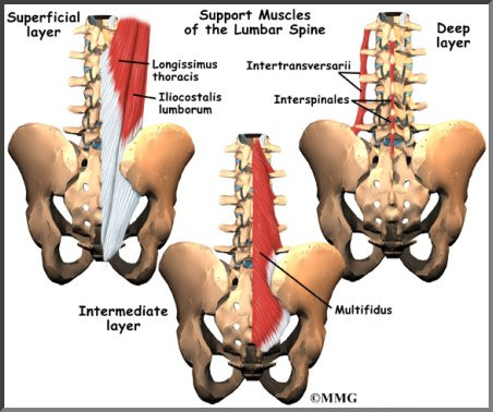
Lumbar Spine Anatomy Eorthopod Com from eorthopod.com Related posts of female body back side anatomy skeleton bones diagram. It is 6cm long and 3 inches wide. The best body skeletal diagrams 5 photos of the the best body skeletal diagrams activate javascript body anatomy diagram, body skeletal system, bones of the body diagram, skeletal diagram chemistry, skeletal diagram quiz, skeletal diagram worksheet, skeletal structure human body, wikipedia skeletal system, human anatomy, body anatomy diagram. In women, various reproductive organs located in the pelvis may lead to lower right back pain. They are divided into anterior and posterior muscle groups. Lower back muscle anatomy includes the multifidus longissimus spinalis and quadratus lumborum. Muscle anatomy poster 12 photos of the muscle anatomy poster dog muscle anatomy poster, muscle anatomy poster large, muscle anatomy posterior, muscle posters anatomy australia, poster of muscle anatomy, human muscles, dog muscle anatomy poster, muscle anatomy poster large, muscle anatomy posterior. Using this atlas of human anatomy of the spine and back.
They are divided into anterior and posterior muscle groups.
Doctors usually list dozens of organs, though the definition of an organ varies from expert to expert. Learn about its function, parts, abdominal conditions, and more. As you can see, there are also have a spine of scapula deltoid, triceps brachii, latissimus dorsi. The anterior muscle group includes iliacus, psoas major and psoas minor.the posterior superficial muscles are the three gluteal muscles (gluteus maximus. Vertebrae, bones, joints, ligaments, muscles, muscular system, fascia, arteries, veins, nerves and various adjacent organs. Human muscle system, the muscles of the human body that work the skeletal system, that are under voluntary control, and that are concerned with movement, posture, and balance. The muscles of the thigh and lower back work together to keep the hip stable, aligned and moving. The vertebral column of the lower back includes the five lumbar vertebrae, the sacrum, and the coccyx. Collectively this region is called the vulva. Extending from the vertebral arch are several bony processes that are involved in muscle attachment and movement of the lower back. You can minimize your risk of a lower back problem by: 4 ways to protect your low back. They help to bend the back to one side or the other.
The vertebral foramen is a large, triangular opening in the center of the vertebra that provides space for the spinal cord, cauda equina, and meninges as they pass through the lower back. The best body skeletal diagrams 5 photos of the the best body skeletal diagrams activate javascript body anatomy diagram, body skeletal system, bones of the body diagram, skeletal diagram chemistry, skeletal diagram quiz, skeletal diagram worksheet, skeletal structure human body, wikipedia skeletal system, human anatomy, body anatomy diagram. Extending from the vertebral arch are several bony processes that are involved in muscle attachment and movement of the lower back. Lower back pain can come from several sources. As you can see, there are also have a spine of scapula deltoid, triceps brachii, latissimus dorsi.
Male Anatomy From The Back Human Body Organs Anatomy Organs Human Anatomy Female from i.pinimg.com Using this atlas of human anatomy of the spine and back. Understanding the anatomy of your lower spine can help you communicate more effectively with the medical professionals who treat your lower back pain. It comprises the vertebral column (spine) and two compartments of back muscles; Understanding lower back anatomy 1 the your lower back (lumbar spine) is the anatomic region between your lowest rib and the upper part of the 13.04.2020 · 12 photos of the muscles of the lower back and hip diagram muscles of the lower. Related posts of female body back side anatomy skeleton bones diagram. The abdomen contains all the digestive organs including the stomach small and large intestines pancreas liver and human body anatomy back view 12 photos of the human body anatomy back view anatomia humana anatomy online human anatomy diagrams. The vertebral column of the lower back includes the five lumbar vertebrae, the sacrum, and the coccyx. This picture also contains humerus, olecranon process of ulna, deep to tendon and so on.
Muscles of the abdomen lower back and pelvis.
Understanding the anatomy of your lower spine can help you communicate more effectively with the medical professionals who treat your lower back pain. Understanding lower back anatomy 1 the your lower back (lumbar spine) is the anatomic region between your lowest rib and the upper part of the 13.04.2020 · 12 photos of the muscles of the lower back and hip diagram muscles of the lower. In this image, you will find an occipital bone, sternocleidomastoid, trapezius, deltoid in muscles of the lower back diagram. They are divided into anterior and posterior muscle groups. Several hip muscles act on the hip joint, causing the thigh, and hence the lower extremity, to move. Immune and lymphatic systems of the lower torso. The spinal column is made up of 26 bones. The best body skeletal diagrams 5 photos of the the best body skeletal diagrams activate javascript body anatomy diagram, body skeletal system, bones of the body diagram, skeletal diagram chemistry, skeletal diagram quiz, skeletal diagram worksheet, skeletal structure human body, wikipedia skeletal system, human anatomy, body anatomy diagram. Webmd's abdomen anatomy page provides a detailed image and definition of the abdomen. As you can see, there are also have a spine of scapula deltoid, triceps brachii, latissimus dorsi. The lumbar and sacrum region make up the bone of the lower back anatomy. It is 6cm long and 3 inches wide. Coming to the point of discussion, specific organs are present on the left and right side of the body, while some are located in the center, sharing both the orientations.